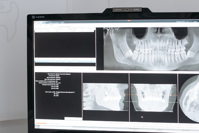
Dental X-Ray and Dental Examination at Basmat Al Hayat Medical Center
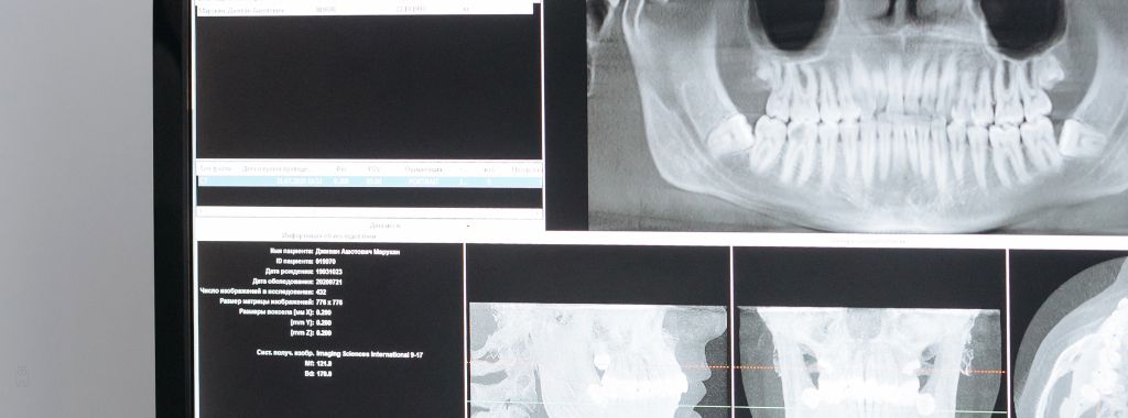
Dental X-Ray and Examination in Muwailih and Ras Al Khaimah
Dental X-rays help the dentist see details that may not be visible during a regular examination inside the mouth. A tooth may look normal from the outside, while there may be decay between the teeth, inflammation near the root, bone loss, an impacted tooth, a problem under an old filling, or changes around a previous restoration. This is why dental X-rays can be an important part of diagnosis and treatment planning in many dental cases.
At Basmat Al Hayat Medical Center, we provide dental X-ray and dental examination services as part of our dental treatment services at our Muwailih – Sharjah and Ras Al Khaimah branches. The visit starts with listening to the patient’s complaint and examining the teeth and gums. The dentist then decides whether an X-ray is needed and which type of image is suitable for the condition. The aim is to use dental imaging when it supports diagnosis and treatment planning, not as a random step for every patient.
This page explains the role of dental X-rays during examination, when they may be needed, what they can help detect, the common types of dental radiographs, and how they support treatments such as fillings, root canal treatment, tooth extraction, dental implants, crowns, bridges, and pediatric dental care. The information is for patient education only. The decision to take an X-ray and choose the type of image is made by the dentist after assessment.
What Is a Dental X-Ray?
A dental X-ray is a diagnostic image that uses a controlled amount of X-ray radiation to show teeth, roots, jawbone, and surrounding structures. Hard tissues such as teeth and bone appear differently from soft tissues, allowing the dentist to evaluate areas that cannot always be seen directly during a normal checkup.
Dental X-rays do not replace a clinical examination. They support it. The dentist still needs to examine the mouth, check the gums, test sensitivity or pain, assess fillings and restorations, and understand the patient’s symptoms. X-rays are used when more information is needed to confirm a diagnosis or plan treatment. In some cases, imaging is needed before treatment can be decided. In other cases, the dentist may not request an X-ray if the problem is clear and imaging is not necessary.
Dental X-rays may be used for different diagnostic and treatment reasons. They can help the dentist check the depth of decay, how close decay is to the nerve, the condition of roots, healing after root canal treatment, wisdom tooth position, bone support, or the area planned for a dental implant. The dentist explains why imaging is needed when it is recommended.
Why Might You Need a Dental X-Ray?
The dentist may request a dental X-ray when more information is needed than what can be seen during a regular examination. Sometimes the patient can point to the painful tooth clearly. Other times, pain may be difficult to locate or may spread across several teeth. X-rays can help the dentist narrow down the possible causes and understand the condition more accurately.
You may need a dental X-ray in situations such as:
- Continuous or repeated tooth pain.
- Strong sensitivity to hot or cold drinks.
- Suspected decay between the teeth.
- Deep decay close to the nerve.
- Pain while chewing or pressing on a tooth.
- Gum swelling or an abscess near a tooth root.
- Assessment before root canal treatment or root canal cleaning.
- Planning simple or surgical tooth extraction.
- Evaluating impacted wisdom teeth or unerupted teeth.
- Planning dental implants or jawbone grafting.
- Monitoring children’s tooth development.
- Checking old fillings, crowns, or bridges.
Having one of these situations does not always mean an X-ray is required, but it means the dentist may consider imaging after examination. The type of X-ray also depends on the goal. The dentist may need a small image of one tooth, a wider image of the jaws, or another type of imaging when the treatment plan is more complex.
What Can Dental X-Rays Help Detect?
Dental X-rays can help the dentist detect or evaluate several issues, including cavities between teeth, decay under old fillings, infection around tooth roots, bone loss related to gum disease, impacted teeth, remaining roots, dental abscesses, and tooth condition before fillings, extraction, implants, crowns, or bridges.
In some cases, decay starts between teeth and cannot be seen easily during a visual examination. X-rays may help detect it before it becomes deeper. In other cases, pain may come from inflammation around the root. The image can help the dentist assess whether there are changes in the bone near the tooth root.
X-rays are also helpful for checking previously treated teeth. If a patient has a large filling, previous root canal treatment, a crown, or a bridge, the dentist may need an image to check the tooth underneath or the surrounding area. This makes dental imaging useful for follow-up, not only for emergency pain.
Common Types of Dental X-Rays
There are different types of dental X-rays. The dentist chooses the suitable type based on the patient’s condition and the information needed. Not every patient needs every type, and the choice is based on diagnosis and treatment planning, not simply on the name of the image.
Small Intraoral X-Rays
Small intraoral X-rays are often used to capture detailed images of one tooth or a small group of teeth. They may help the dentist check decay, roots, bone around the tooth, or root canal treatment. They are commonly used when pain is located in a specific area or before fillings and root canal treatment.
Bitewing X-Rays
Bitewing X-rays may help detect decay between teeth and assess bone levels around back teeth. These areas can be difficult to see with the eye, especially when teeth are close together or when decay is still early.
Periapical X-Rays
A periapical X-ray shows the full tooth, from the crown to the root tip and the surrounding area. It may be useful when there is deep pain, suspected root infection, root canal treatment planning, or assessment before extraction.
Panoramic X-Rays
A panoramic X-ray, when available or needed, gives a wider view of the jaws and teeth. It may be used to assess wisdom teeth, impacted teeth, tooth development, jaw structure, or planning for certain treatments such as extraction or implants. However, a panoramic image may not replace a smaller detailed X-ray when the dentist needs to assess a specific tooth closely.
Additional Imaging When Needed
In more advanced treatment plans, such as dental implants or complex cases, the dentist may need more detailed imaging depending on availability and clinical need. The dentist explains the type of imaging and why it is useful for the case.
Clinical Examination vs. Dental X-Ray
A clinical examination is the direct evaluation that the dentist performs inside the mouth. During this examination, the dentist can see visible decay, gum inflammation, tooth fractures, tooth movement, tartar buildup, or pain when pressing on a tooth. However, the dentist cannot always see what is happening inside the tooth, under the gum, between the teeth, or around the roots.
Dental X-rays complement the examination by showing areas that are not directly visible. They may reveal the depth of decay, the condition of a root, the level of bone support, or the position of an impacted tooth. In many cases, a good diagnosis depends on combining the clinical examination, the patient’s symptoms, medical and dental history, and X-rays when needed.
If the dentist requests an X-ray, it does not mean the examination was not useful. It means the case needs more information before a treatment decision is made. This is especially important for root canal treatment, surgical extraction, dental implants, and unclear dental pain.
Are Dental X-Rays Safe?
Dental X-rays use a low amount of radiation when performed properly, but they should still be used only when needed and based on the dentist’s decision. The dentist weighs the diagnostic benefit against radiation exposure and aims to keep exposure as low as reasonably achievable. This is especially important for children, pregnant patients, or patients who need repeated imaging.
If you are pregnant, think you may be pregnant, or have concerns about X-rays, you should inform the dentist before imaging. The dentist will decide whether the X-ray is necessary at that time, whether it can be delayed, and what precautions are suitable for the situation.
For children, dental X-rays are requested only when needed. This may include checking tooth development, detecting hidden decay, assessing an injury, or understanding the position of permanent teeth. X-rays should not be used for children without a clear reason. The dentist chooses the appropriate type based on the child’s age and condition.
How Is a Dental X-Ray Taken?
The process depends on the type of image needed. For small intraoral X-rays, a small sensor or film is placed inside the mouth near the tooth being examined. The patient is asked to stay still for a few seconds while the image is taken. For wider images such as panoramic imaging, the patient stands or sits in a specific position while the machine moves around the head, depending on the device.
Before imaging, the team may ask the patient to remove metal items that may affect the image, such as certain jewelry near the face or mouth. The patient is guided on how to stand, bite, or stay still. Movement can make the image unclear and may require repeating the image, so proper positioning is important.
After the image is taken, the dentist reviews it and explains the findings to the patient in a simple way. In some cases, one image is enough to plan treatment. In other cases, the dentist may need an additional examination or another image if the case is more complex.
Dental X-Rays Before Fillings
Before dental fillings, the dentist may request an X-ray if decay is not clearly visible, is located between the teeth, or appears deep. The X-ray helps the dentist understand how far the decay has spread and whether it is close to the nerve. If decay is limited, a filling may be enough. If it has reached the nerve or is very close to it, root canal treatment or a different treatment plan may be needed.
This assessment helps reduce surprises during treatment. A tooth may look suitable for a filling from the outside, but an X-ray may show that the damage is deeper than expected. The dentist uses both the examination and the image to explain the treatment plan.
Dental X-Rays Before Root Canal Treatment
In root canal treatment, X-rays are often important because they help the dentist assess root length, expected canal shape, signs of infection around the root tip, and the condition of any previous root canal treatment. The dentist may need an image before treatment and sometimes during or after treatment depending on the case.
Root canal cleaning requires precision because canals differ in shape and number from one tooth to another. X-rays help the dentist understand root direction and evaluate changes around the tooth. However, the treatment plan is not based on X-rays alone. Symptoms, clinical testing, and examination are also considered.
Dental X-Rays Before Tooth Extraction
Before simple or surgical tooth extraction, the dentist may need an X-ray to assess root shape, tooth position, surrounding bone, infection around the root, or the position of a wisdom tooth. This is especially important for back teeth, broken teeth, and impacted wisdom teeth.
Imaging helps the dentist determine whether the tooth may be suitable for simple extraction or whether surgical extraction may be needed. It also helps the dentist explain the treatment plan and aftercare instructions to the patient before starting.
Dental X-Rays for Dental Implants and Jawbone Grafting
Dental implant treatment requires careful assessment of the jawbone. X-rays or imaging may be an important part of planning because they help evaluate bone height, width, density, and the location of nearby structures such as nerves or sinus areas depending on the implant location. If bone support is not enough, the dentist may discuss jawbone grafting or other tooth replacement options.
Imaging alone does not decide whether implants are suitable. The dentist also evaluates gum health, neighboring teeth, general health, and bite. All of these factors are considered before creating the final treatment plan.
Dental X-Rays for Children
Children may need dental X-rays in selected cases, such as hidden decay, monitoring permanent teeth, assessing dental injury, or understanding why a tooth has not erupted. Because children’s teeth change as they grow, imaging may help the dentist understand the relationship between baby teeth and permanent teeth when needed.
At Basmat Al Hayat Medical Center, dental X-rays for children are requested based on need, and the reason is explained to parents. X-rays may support treatment planning before baby tooth decay treatment, fluoride protection, or pediatric dental care when the dentist needs more information than a visual exam can provide.
Do You Need Dental X-Rays at Every Visit?
No. Patients do not need dental X-rays at every visit. The need depends on age, symptoms, history of decay, pain, type of treatment required, and whether the dentist is monitoring a previous condition. If the patient has recent clear dental X-rays from another clinic, the dentist may be able to review them instead of repeating imaging, depending on their quality and relevance.
It is helpful to tell the dentist if you have had recent dental X-rays elsewhere. The dentist may review them if they are available and decide whether they are enough or whether a newer or different image is needed.
How to Prepare for a Dental X-Ray
Most dental X-rays do not require special preparation. However, it is important to inform the dentist or team if you are pregnant or think you may be pregnant, if you have difficulty opening your mouth, if you have a strong gag reflex when something is placed inside your mouth, or if you have recent dental X-rays.
For intraoral X-rays, you may feel mild discomfort from the sensor or film inside the mouth, but it usually takes only a short time. If you feel nervous, tell the team so they can guide you calmly. The goal is to obtain a clear image the first time whenever possible.
What Happens After the X-Ray?
After the X-ray is taken, the dentist reviews the image together with the clinical examination. The dentist may explain the location of decay, the condition of the root, the reason for pain, or why a specific treatment is recommended. In some cases, the image confirms that the tooth needs a filling only. In other cases, it may show the need for root canal treatment, extraction, a crown, a bridge, an implant, or monitoring without immediate treatment.
If the X-ray does not clearly show the cause of pain, the dentist may need additional tests or follow-up. Not all dental pain appears clearly on an X-ray. This is why diagnosis should combine symptoms, examination, imaging when needed, and patient history.
Common Misunderstandings About Dental X-Rays
One common misunderstanding is that X-rays show everything by themselves. In reality, X-rays are important diagnostic tools, but they are not a replacement for the dentist’s examination. The dentist may still need to check the gums, test sensitivity, evaluate the bite, and understand the patient’s symptoms to reach a more accurate diagnosis.
Another misunderstanding is that X-rays are needed at every visit. Proper use of dental X-rays depends on clinical need, not routine use without a reason. On the other hand, avoiding X-rays when they are needed may delay diagnosis, especially in cases of deep pain, infection, surgical extraction planning, or dental implant assessment.
Dental X-Ray as Part of Dental Treatment Services
Dental X-rays are connected to many dental treatment services. They may be part of decay diagnosis before fillings, nerve assessment before root canal treatment, planning for simple or surgical extraction, checking jawbone before dental implants, or monitoring children’s teeth. This makes dental imaging an important step in the patient journey from symptoms to diagnosis and treatment.
Internally linking this page to dental fillings, root canal treatment, root canal cleaning, tooth extraction, dental implants, jawbone grafting, and pediatric dental treatment helps patients understand why imaging may be needed for different services. It also helps the website build a clear and useful service structure.
Why Choose Basmat Al Hayat Medical Center for Dental X-Ray and Examination?
At Basmat Al Hayat Medical Center, dental examination starts with understanding the patient’s concern and assessing the condition. Dental X-rays are requested when they are needed to support diagnosis or treatment planning. The dentist explains why imaging is being requested and what they are looking for in the image, then connects the findings to a suitable treatment plan.
Dental X-ray and examination services are available as part of our dental treatment services at our Muwailih – Sharjah and Ras Al Khaimah branches. Having services such as dental fillings, root canal treatment, extraction, crowns, bridges, dental implants, and pediatric dental care available in the center helps patients continue their treatment plan when care is needed after diagnosis.
Basmat Al Hayat Medical Center Branches
Muwailih Branch – Sharjah
Sheikh Khalifa Street, Muwailih Commercial
Customer Service: 065597444
Reception: 0544449500
Working Hours: Saturday to Thursday, 9:00 AM to 9:00 PM
Ras Al Khaimah Branch
Al Dhait Area – Agencies Street
Customer Service: 072222256
Reception: 0506462004
Working Hours: Saturday to Thursday, 9:00 AM to 9:00 PM
Friday: 12:00 PM to 8:00 PM
Booking a Dental X-Ray and Examination Appointment
If you have tooth pain, strong sensitivity, suspected hidden decay, gum swelling, or need assessment before dental treatment, you can contact Basmat Al Hayat Medical Center to arrange an examination at our Muwailih or Ras Al Khaimah branch. After the checkup, the dentist will decide whether a dental X-ray is needed and which imaging type is suitable for your condition.
Dental X-Ray FAQ
Are dental X-rays needed for every patient?
No. Dental X-rays are requested based on clinical need and examination findings. Some patients need imaging for diagnosis or treatment planning, while others may not need it during the same visit.
Are dental X-rays painful?
The X-ray itself is not painful. Some patients may feel mild discomfort when a small sensor is placed inside the mouth, but the process usually takes a short time.
What can dental X-rays show?
They may help detect decay between teeth, root inflammation, bone loss, impacted teeth, remaining roots, and tooth condition before fillings, root canal treatment, extraction, or implants.
Can children have dental X-rays?
Yes, when needed. The dentist may request X-rays for children to check hidden decay, permanent tooth development, injuries, or other dental concerns. The type is chosen based on age and condition.
Can dental X-rays be taken during pregnancy?
You should tell the dentist if you are pregnant or think you may be pregnant. The dentist will decide whether the X-ray is necessary, whether it can be delayed, and what precautions are appropriate.
Do X-rays replace a dental examination?
No. X-rays support the examination but do not replace it. The dentist combines symptoms, clinical examination, and imaging when needed for better diagnosis.
How often do I need dental X-rays?
There is no single schedule for everyone. It depends on age, dental condition, history of decay, pain, treatment needs, and the dentist’s assessment.